Pixel classification with Machine learning
Z-cells is inspired from hyperspectral imaging. It does not rely (only) on the in-focus image, but use the information contained in several focal planes (z-stack). This allows to identify cellular region with high fidelity without making assumption on the cells morphologies. The central idea is that cells contour, cells interior or any objects within the field of view have different intensity profiles in the z direction. Using machine learning, it is possible to train an algorithm to classify each pixels based on their focal signature. The method is very robust and gives excellent results on E. coli (rod shaped), S. cerevisiae (round shape), mammalian epithelial (HeLa) cells. The method is even reliable on a mixture of bacteria and yeast cells.

Precise results
The current method is still in development but already gives excellent results (less than 1% classification errors) with as few as ~ 10 images in the z-stack. It can be done with a conventional automated microscope, or with a dedicated piezo controller to improve the reliability of the z-stack acquisition. This usually improves classification performance.

For any cell Types
With a proper training dataset, cells can be detected and classified irrespective of their morphologies. The method can be used with bacteria (rod shaped), yeast (round) or mamalian cells (complex shapes). In this preliminary version, a simple matlab Graphical User Interface is provided to help with training the machine learning algorithm.

Modular
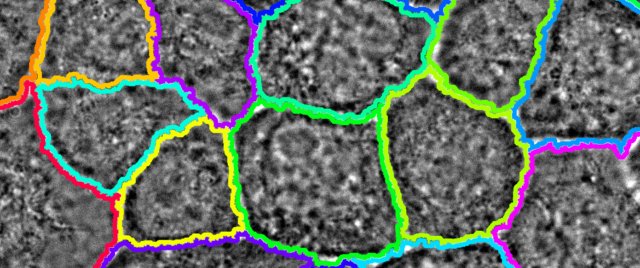
The output of the stack analysis is a classification map of the pixels. This allows to get a very good segmentation with simple methods (watershed), but it can be plugged as the entry of more complex methods to get an even better segmentation and tracking of cells in the picture.
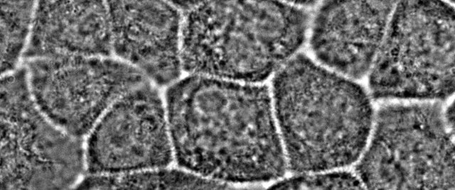
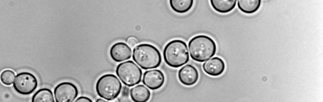
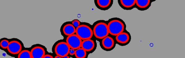
Classification examples
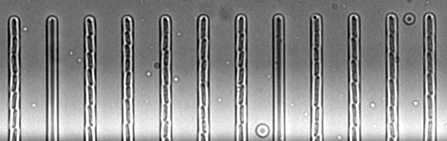
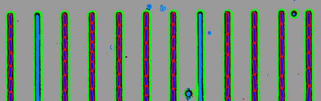
Use the slider to change from the original in focus image to the result of the classification. For HeLa cells, we actually show segmentation results based on the classification. Classifications were obtained with a IX71 microscope equipped with a PI piezzo controler and with bright field illumination.
Bacteria | E. Coli
Yeast | S. Cerevisiae
Mamalian | HeLa cells